iPhone Ultrasound Probe Aids in Life-Saving Aortic Diagnosis
A portable iPhone ultrasound probe enabled doctors to diagnose an aortic dissection in time, saving a patient’s life at Robert Wood Johnson University Hospital.
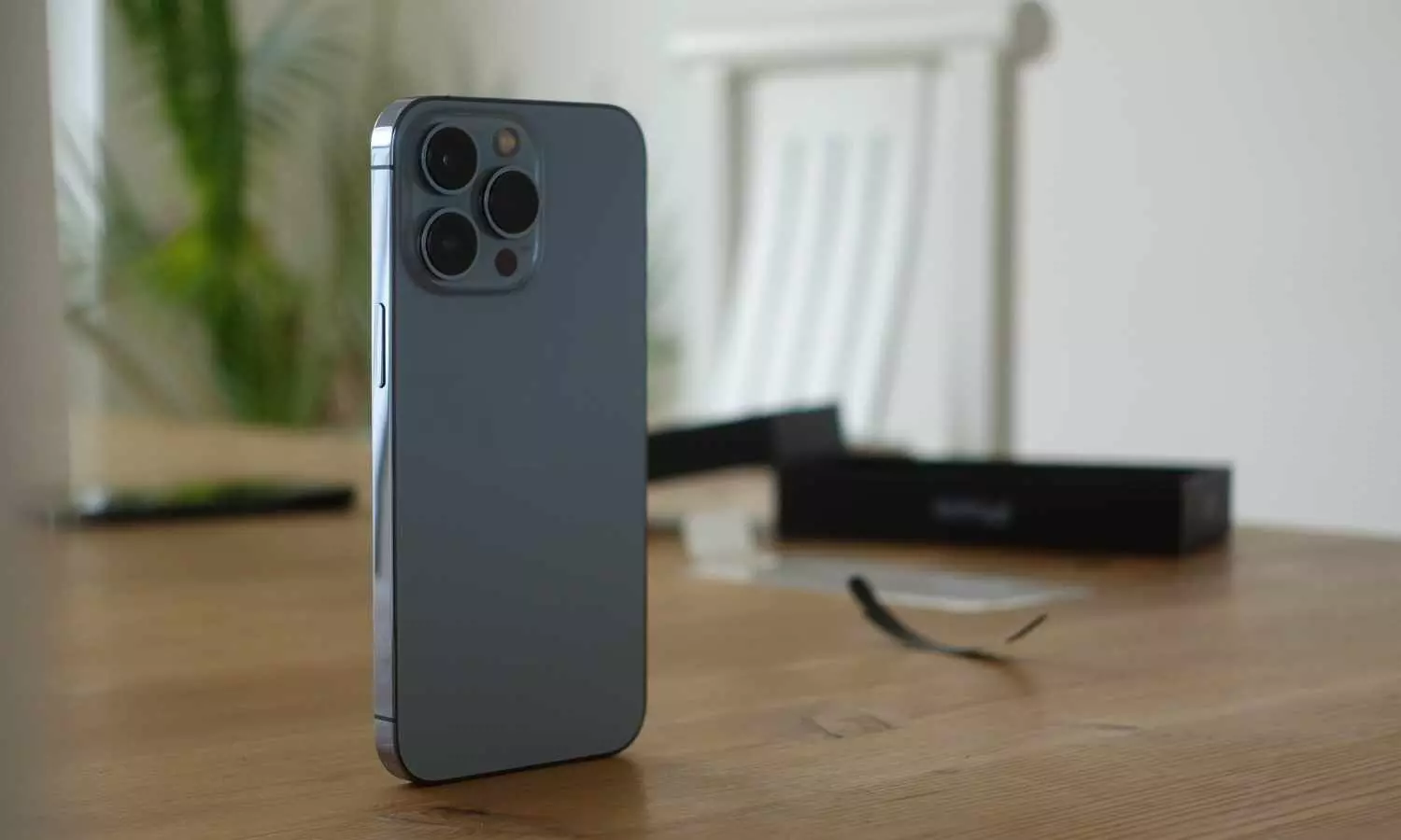
image for illustrative purpose

A groundbreaking handheld ultrasound device connected to an iPhone proved crucial in diagnosing a critical cardiac condition, enabling life-saving surgery for a hospital analyst who suffered an aortic dissection. The real-time imaging technology, used at Robert Wood Johnson University Hospital, expedited treatment and prevented a fatal outcome.
Sara Adair, a mother of two with a family history of aortic dissection, experienced severe chest pain on July 22, 2024. Diagnosed with Loeys-Dietz syndrome, a genetic disorder affecting connective tissue, she underwent regular check-ups, yet no warning signs of an impending cardiac emergency had been detected. The pain escalated and radiated to her neck, a known symptom of an aortic tear. Before she could alert her husband, she collapsed.
Emergency responders transported Adair to the hospital, where initial assessments pointed to a possible heart attack. This common misdiagnosis could have been fatal, as treatments for heart attacks differ significantly from those for aortic dissections. Without prompt surgical intervention, the survival rate decreases by up to 2 per cent per hour.
In the emergency department, Dr. Shawn Chawla, a cardiology fellow, utilized an iPhone-compatible handheld ultrasound probe to conduct a rapid scan. The portable device provided immediate imaging, revealing a significant tear in Adair’s aorta. Further diagnostic evaluations confirmed the severity of the condition, prompting an urgent open-heart surgery.
Dr. Partho P. Sengupta, chief of cardiology at the hospital, highlighted the role of handheld ultrasound in critical care. “More than half of patients with aortic dissections do not reach the hospital in time. Early detection is essential for survival,” he stated. The iPhone ultrasound probe enabled a swift and accurate diagnosis, facilitating immediate surgical intervention.
Cardiac surgeon Dr. Hirohisa Ikegami led the complex open-heart procedure. While Adair faced complications, including fluid buildup and a stroke during surgery, she has been recovering through cardiac rehabilitation. The integration of smartphone-based ultrasound technology into emergency medicine has demonstrated its potential to transform diagnostic protocols and improve patient outcomes.
Reflecting on her experience, Adair credited the advanced medical imaging technology for her survival. “If I had been misdiagnosed with a heart attack, I might not have survived,” she said.
With a history of aortic dissection in her family, Adair’s children are now undergoing genetic testing for Loeys-Dietz syndrome.

